Survey One 2002
Supplied CD68
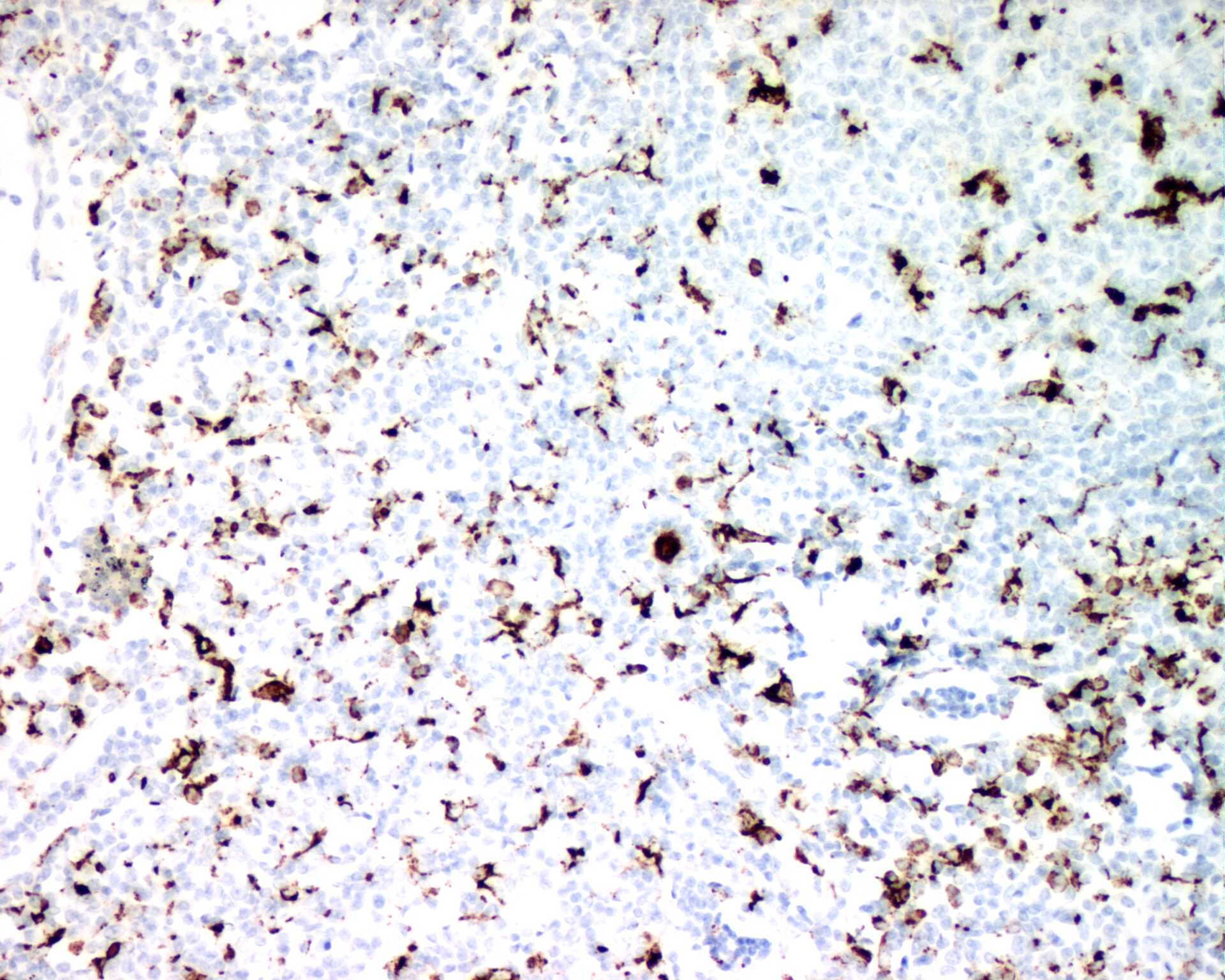
A section of a reactive lymph
node shows follicular hyperplasia, histocytosis and focal increase in vascularity. The CD68 antigen is
mainly located in lysosomes. It belongs to a family of plasma membrane shuttling proteins which play a
role in endocytosis and/or lysosomal traffic. The antibody from clone PG-M1 stains macrophages in
spleen, gut, lung and bone marrow. It also stains Kupffer's cells but not granulocytes and their
precursor cells. Plasmacytoid T cells, which are present in many reactive lymph nodes and are believed
to be of monocyte/macrophage origin, are positive as well. The antibody helps in differentiation of
acute myelomonocytic (M4) and monocytic (M5) leukaemias from other types of acute myeloid leukaemias
and chronic myeloid leukaemia.
The best result is obtained by peroxidase blocking for 10 min., pressure cooking treatment for 2.5
min., and immuno-stained with 1:50 diluted primary antibody for 32 min. at 37 oC. |
 |
References:
Falini B, et al. PG-M1: a new monoclonal antibody directed against a fixative-resistant epitope on the
macrophage-restricted form of the CD68 molecule. Am J Pathol 1993; 142:1359-72.
Holness CI, Simmons DL. Molecular cloining of CD68, a human macrophage marker related to lysosomal
glycoproteins. Blood 1993; 81:1607-13.
Fukuda M. Lysosomal membrane glycoproteins. Structure, biosynthesis, and intracellular trafficking. J Biol
Chem 1991; 266:21327-30.
S100
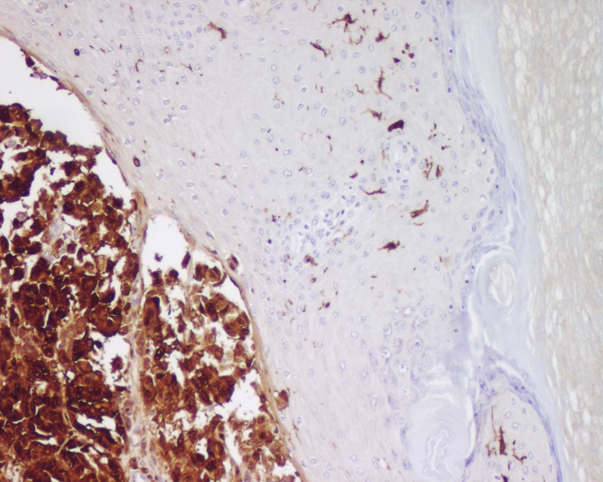
A section of a melanoma shows
diffuse and strong immuno-staining in both the nucleus and cytoplasm. In the areas not shown here,
S100 also stains Langerhans cells, skin appendage structure, melanocytic cells, nerve bundles and
lipocytes. It also labels glial and ependymal cells in brain, Schwann cells in peripheral nervous
system, and interdigitating reticulum cells in lymph nodes. It can be used to distinguish tumours
derived from the aforesaid cells versus other cell origins. In addition, it is useful for recognizing
tumours of salivary glands and cartilage.
The best result is obtained by peroxidase blocking for 20 min., trypsination for 6 min., and
immuno-stained with 1:1000 diluted primary antibody for 32 min. at 37 oC. There is
suggestion for reducing background staining by pretreatment with 2% Tween 20 and incorporation
of 2% Tween 20 in the immunological reagents. |
 |
References:
Moore BW. A soluble portein characteristic of the nervous system. Biochem Biophys Res Comm 1965;
19:739-44.
Kindblom L-G, et al. S-100 protein in melanocytic tumors. Acta Path Microbiol Immunol Scand Sect A
1984; 92:219-30.
Wick MR, et al. Recognition of malignant melanoma by monoclonal antibody HMB-45. An immunohistochemical
study of 200 paraffin-embedded cutaneous tumors. J Cutan Pathol 1988; 15:201-7.
Juhl BR, et al. The effect of Tween 20 on indirect immunoperoxidase staining of blood group antigen A
in human urothelium. J Histochem Cytochem 1984; 32:935-41.

Last updated on 3 November, 2002.
Prepared by HKIMLSQAP Anatomical Pathology Panel.
Copyright 2001-2002 HKIMLSQAP. All Rights Reserved.
