Survey Three 2002

Supplied Myeloperoxidase

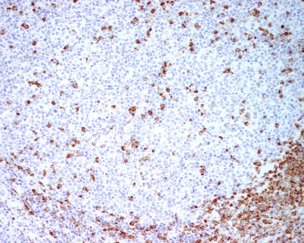
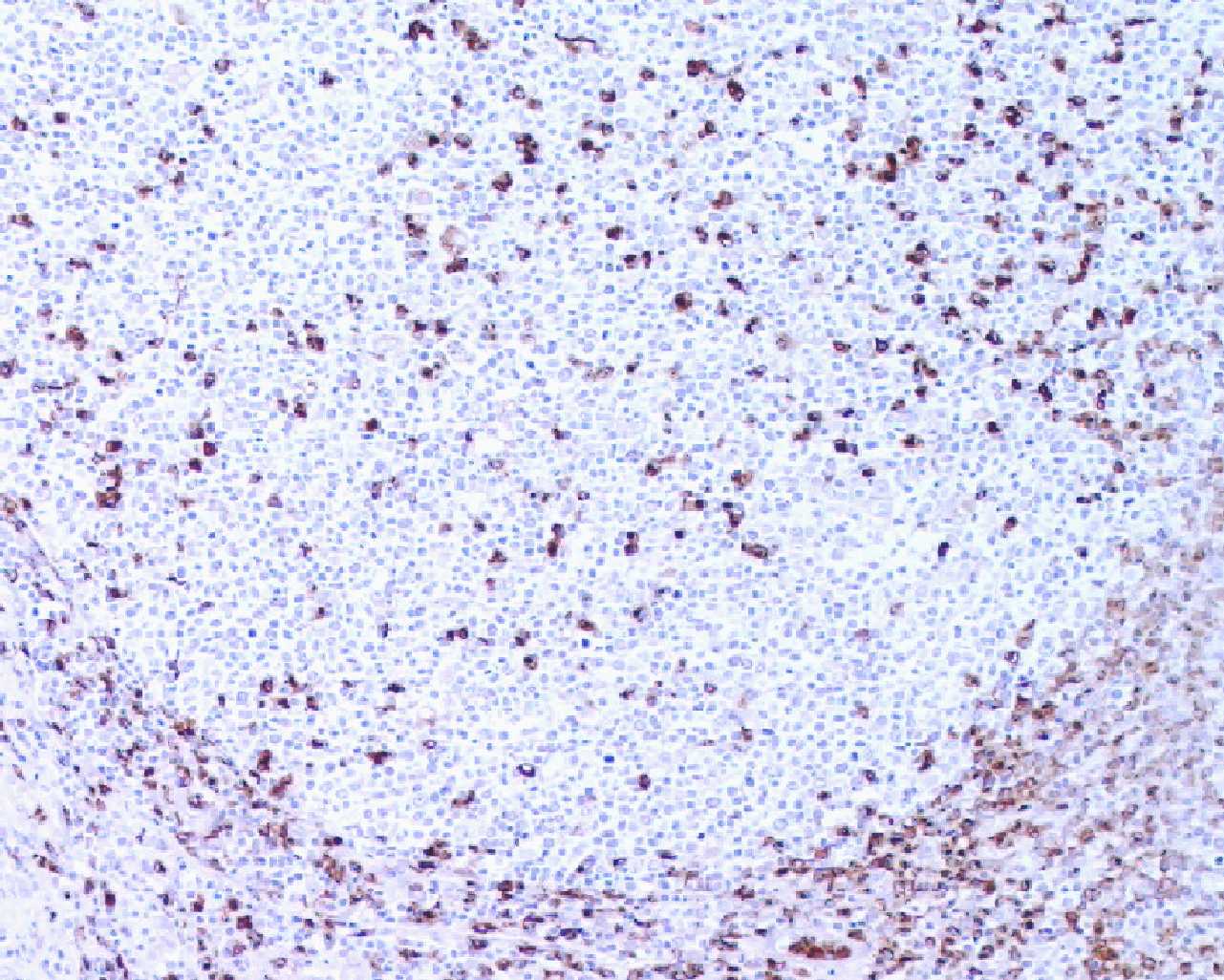
The micrograph is a reactive lymph node with follicular hyperplasia, sinus histiocytosis and focal increase in vascularity. The anti-myeloperoxidase antibody may be used to discriminate between lymphoid leukaemias and myeloid leukaemias.

The best results is obtained by peroxidase blocking for 10 min., microwave irradiation for 15 min., and immuno-stained with 1:500 diluted primary antibody for 60 min. at room temperature.


In-house Myeloperoxidase

A section from same tissue block is immunostained with in-house myeloperoxidase antibody. The average result is better than that stained with supplied antibody. The best result is obtained by peroxidase blocking for 10 min., pressure cooking treatment for 2.5 min., and immuno-stained with 1:1500 diluted primary antibody for 32 min. at 37 oC.


Last updated on 9 November, 2002.

Prepared by HKIMLSQAP Anatomical Pathology Panel.

Copyright 2001-2002 HKIMLSQAP. All Rights Reserved.